Western Blotting Allows The Detection Of Specific Proteins Within A Complex Biological Sample
 |
| Western Blotting |
Western
Blotting, also known as immunoblotting, is a powerful and
widely used laboratory technique that allows the detection and characterization
of specific proteins within a complex biological sample. This technique plays a
crucial role in molecular biology, cell biology, and biochemistry research,
enabling scientists to study protein expression levels, post-translational
modifications, and protein-protein interactions.
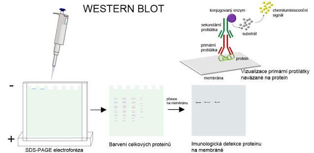
The blotting process
involves several key steps. First, the sample containing the proteins of
interest is separated by gel electrophoresis. Typically, sodium dodecyl
sulfate-polyacrylamide gel electrophoresis (SDS-PAGE) is employed, which
separates the proteins based on their size. The separated proteins are then
transferred (blotted) from the gel to a solid membrane, usually made of
nitrocellulose or polyvinylidene fluoride (PVDF). This transfer step is crucial
as it allows for better protein visualization and interaction with antibodies.
According
To Coherent Market Insights, The
Western Blotting Market Is Anticipated To Reach A Value Of
US$ 759.5 Million In 2023 And Is Projected To Grow At A 6.8% CAGR From 2023 To
2030.
After the transfer in Western Blotting process, the membrane
is blocked with a protein-rich solution (often containing milk or bovine serum
albumin) to prevent nonspecific binding of antibodies and to minimize
background signals. The membrane is then probed with primary antibodies that
specifically target the protein of interest. These antibodies bind to their
respective target proteins on the membrane, forming an antigen-antibody
complex.
Next, the membrane is
washed to remove any unbound primary antibodies. In some cases, a secondary
antibody labeled with an enzyme or a fluorescent marker is added. This
secondary antibody recognizes and binds to the primary antibody, amplifying the
signal and enhancing detection sensitivity. Alternatively, direct detection
methods utilizing labeled primary antibodies can be used, omitting the need for
a secondary antibody.
The final step in the Western Blotting process is the
visualization of the protein bands. This can be achieved using various methods,
such as chemiluminescence or fluorescence. In chemiluminescence, an enzyme
attached to the secondary antibody catalyzes a reaction that produces light,
which is then captured on an X-ray film or detected with a specialized imaging
system. Fluorescently labeled antibodies, on the other hand, emit fluorescence
when excited by specific wavelengths of light, which can be visualized using
appropriate imaging equipment.
Pleural Diseases
are a set of pathological conditions originating in the pleural cavity,
encompassing diseases like pleural plaques caused by asbestos exposure, pleural
thickening, and malignant pleural effusions resulting from cancerous growth in
the pleura. These conditions can significantly affect breathing and necessitate
specialized medical management.
Western
Blotting offers several advantages, including its versatility
and sensitivity. It allows researchers to examine multiple proteins
simultaneously within a single sample and enables the detection of low
abundance proteins. Moreover, by using specific antibodies, researchers can
gain insights into protein isoforms, modified forms, and protein-protein
interactions.


Comments
Post a Comment